Q4. What are critical points to get better survival of embryos?
Following points should be considered.
1. To minimize contamination,
We have performed in vivo electroporation in a clean room (SPF mouse facility). To be SPF may not be necessary, but it will be better to clean your bench before surgery. Tools such as forceps and scissors should be sterilized.
2. Gentle and careful manipulation is required.
a. Damages to the placenta and blood vessels should be minimized (do not pinch them with forceps, and do not hurt blood vessels by dragging them on gauze).
b. Exposed tissues, such as the uterus and the yolk sac, should be always kept wet with warm saline (prewarmed at 37°C).
c. It may be better to check the survival rate of embryos two days after just taking out and repositioning the uterus without injection of DNA and electroporation. The survival rate must be 100% or very close to 100%. If the rate is not good enough, some basic procedures of surgery are not good. Check our procedures again sentence by sentence.
3. Leak of electric pulses should be minimized.
Electric pulses should not be delivered to the embryonic heart and placenta as much as possible. The fasciculation of the pregnant mouse by electric pulses is not a good sign, either. Be sure not to touch the electrodes with other tissues of the pregnant mouse, such as the intestine. If you still find the fasciculation, check whether the strength of your pulses is correct and whether your electrodes are correctly insulated.
4. To aviod adhesion of the uterus (or yolk sac),
Fill the abdominal cavity with warm saline before sewing .
5. The surgery from cutting to sewing the skin should be finished within 45 minutes. If it takes longer than 45 minutes to operate all embryos, the number of operated embryos should be reduced.
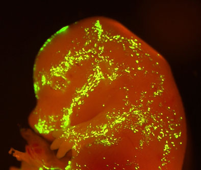
Q5. Transfection efficiency is not good.
The major cause for no expression of genes will be failure of DNA injection. You should practice injection of DNA using Indigo Carmine dye and master where and how to inject the pipette. An atlas of the mouse brain is very helpful to figure out the site of the ventricle. The use of the dye is only for practice. When we perform in vivo electroporation, we do not use the dye to avoid any side effects.
Q6. Which electroporator can be used for in vivo electroporation?
Electroporators that deliver square pulses can be used for in vivo electroporation. Initially we used ElectroSquarePorator T820 (BTX, San Diego, CA), because only the square pulse electroporator was commercially available. After we established the mouse in vivo electroporation system, other electroporators, such as CUY21EDIT (Nepa Gene, Ichikawa, Japan), became available.
Q7. Can we induce gene expression using in vivo electroporation?
Yes. Gene expression is strictly induced in postmitotic neurons after in vivo electroporation, using the tetracycline (Tet)-controlled gene regulatory (Tet-On) system (Sato et al., J. Neurosci. Methods 214, 170-176 (2013)).